Ralpharia magnifica Watson, 1980

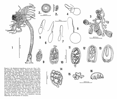
Figures. 1-16. Ralpharia magnifica Watson, 1980. Fig. 1. Fertile hydranth, holotype colony, drawn from underwater photograph. Fig. 2A. Transverse section through mid-stem region showing longitudinal circumferential canals. Fig. 2B. Canals, enlarged. Fig. 3. Blastostyle with gonophores in various stages of development, drawn from underwater photograph. Figs 4-8. Nematocysts (all drawn to same scale). Figs 4-5. Stenoteles from hydranth: Fig. 6. Isorhiza from hydranth: Fig. 7. ? Isorhiza from gonophore. Fig. 8. Anisorhiza from gonophore. Figs. 9-13. Stages in development of the female gonophore (all drawings to same scale from living material): Fig. 9. Immature gonophore. Fig. 10. Nearly mature gonophore with segmented ova: Fig. 11. Free medusa liberating ova. Fig. 12. Later stage, showing nearly spent medusa with few remaining ova. Fig. 13. Empty medusa showing spadix. Fig. 14. Mature ova dissected out from gonophore, showing thin pellicle covering ova (preserved material). Figs. 15-16. metamorphosis of the egg: Fig. 16. Larva 1-2 hours after expulsion, showing beginning of development of tentacles.
From Watson, J.E. (1980) The identity of two tubularian hydroids from Australia with a description and observations on the reproduction of Ralpharia magnifica gen. et sp. nov. Memoirs of the National Museum of Victoria, Melbourne 41: 53-63.
Photographer: Watson, Jeanette. Publisher: Watson, Jeanette.
